


Category
|
Order Code |
Product Name |
|
A-06-02-M03 |
Veterinary Palm B/W Ultrasound Linear Probe | Wireless Linear Array |

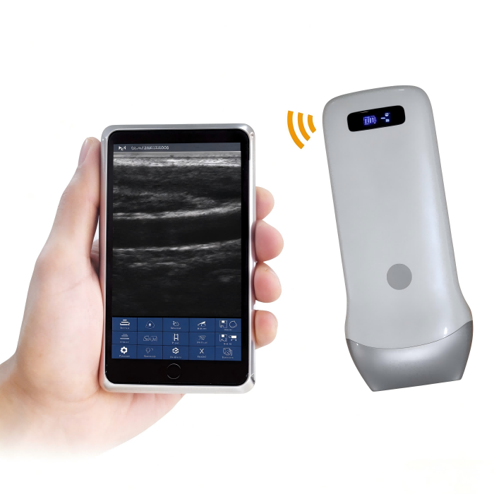
The FKNMED Veterinary Palm B/W Ultrasound Linear is a compact, wireless linear array ultrasound probe designed for veterinary clinical and laboratory applications. It features an 80-element electronic linear array transducer with 7.5/10MHz operating frequencies and connects to compatible smartphones, tablets, or Windows mobile devices to provide B/W ultrasound imaging without a conventional cart or dedicated console.
With a net weight of 250 grams and dimensions of 156mm × 60mm × 20mm, the probe is suitable for mobile examinations in veterinary clinics, animal hospitals, farms, and field environments. Its portable design supports point-of-care imaging where a conventional cart-based ultrasound system may be impractical.
Supports routine small-animal examinations and preliminary imaging assessments in veterinary clinics, pet hospitals, and animal healthcare facilities.
Provides a compact imaging option for intraoperative localization and soft-tissue assessment where portable ultrasound equipment is preferred.
Can be used for post-operative monitoring, fluid assessment, and follow-up examinations without requiring patients to be moved to a separate imaging room.
Supports abdominal, musculoskeletal, and superficial structure assessment within the adjustable 40–100mm scanning depth.
The 10MHz frequency option supports high-frequency linear imaging for superficial and shallow structures in relevant veterinary examination workflows.
Suitable for on-site reproductive and obstetric assessment in livestock and poultry operations, with built-in obstetric measurement functions.
Provides portable imaging support for veterinary laboratories and third-party testing institutions that require mobile image acquisition and examination records.
The 80-element electronic linear array transducer operates at 7.5/10MHz. It provides 256-level gray scale imaging, adjustable noise reduction, and a dynamic range of 40–110 to support image observation across different examination conditions.
The built-in 4200mAh lithium battery provides approximately 3 hours of working time. Its palm-sized design supports examinations in clinics, treatment rooms, farms, and field environments without continuous mains power connection.
Images and videos can be stored directly on connected smartphones or tablets, supporting digital image review and record management without film or printing.
The device supports distance, area, and obstetric measurements. These functions assist examination workflows and provide measurement information for clinical review and reporting.
The probe supports iOS, Android, and Windows platforms, allowing veterinary users to connect it to compatible mobile devices already used in their clinics or laboratories.
The wireless probe configuration avoids the need for a dedicated ultrasound cart or fixed installation. Its portable structure is suitable for veterinary clinics, animal hospitals, farms, and distributors supplying mobile imaging equipment.
| Item | Specification |
|---|---|
| Scan Mode | Electronic Array, Electronic linear array scanning |
| Frequency | Linear probe 7.5 / 10MHz |
| Elements | 80 elements |
| Scanning Depth | Linear 40–100mm, adjustable |
| Field of View | Linear L40 |
| Screen | Smartphone or tablet screen |
| Supporting System | iOS, Android, Windows |
| Display Mode | B, B/M |
| Image Gray Scale | 256 level |
| Noise Reduction | 0–1–2–3–4 |
| Gain | 30dB–105dB |
| Dynamic Range | 40 / 50 / 60 / 70 / 80 / 90 / 100 / 110 |
| Image / Video Storage | Store on mobile phones, Tablet PC |
| Measurement | Distance, area, obstetrics and other |
| Power | Built-in lithium battery, 4200mAh |
| Battery Working Time | 3 hours |
| Size | 156mm × 60mm × 20mm |
| Net Weight | 250 gram |
FKNMED supplies veterinary equipment for animal hospitals, veterinary clinics, farms, laboratories, and international distributors. This wireless linear ultrasound probe offers a portable imaging configuration for buyers seeking mobile examination equipment without the footprint of a conventional cart-based system.
Its compatibility with iOS, Android, and Windows devices, together with built-in measurement functions and mobile image storage, supports integration into different veterinary examination environments. The compact design also provides distributors and importers with an alternative product configuration for their animal imaging equipment portfolios.
For B2B procurement, FKNMED supports inquiries from veterinary equipment distributors, importers, traders, clinics, and institutional buyers. Contact us to discuss product specifications, device compatibility, and supply requirements.
Q1. How does a 7.5/10 MHz palm linear ultrasound probe improve superficial tissue imaging in small animal clinics?
A 7.5/10 MHz linear ultrasound probe supports high-frequency imaging of superficial anatomical structures in small animals, including tendons, ligaments, muscles, skin layers, and peripheral soft tissues. The adjustable frequency allows veterinarians to select an appropriate imaging setting according to the examination target and required penetration depth. In canine and feline practice, this capability is useful for musculoskeletal assessments, localized soft tissue examinations, and superficial lesion evaluation. Its handheld design also allows examinations to be performed directly in consultation rooms or treatment areas, reducing dependence on fixed ultrasound consoles. This makes it a practical option for clinics requiring accessible imaging capabilities within routine diagnostic workflows.\
Q2. What factors should veterinarians consider when selecting an 80-element linear ultrasound probe for feline and canine diagnostics?
When selecting an 80-element linear ultrasound probe for feline and canine diagnostics, veterinarians should evaluate imaging frequency, scanning depth, image resolution, probe design, and software functionality. A 7.5/10 MHz frequency configuration is relevant for superficial tissue examinations, while adjustable scanning depth from 40 mm to 100 mm provides flexibility for different anatomical targets. The 256-level grayscale display supports differentiation of tissue structures, and adjustable gain and dynamic range settings allow operators to optimize image presentation. Compatibility with the clinic's existing mobile devices, battery runtime, and image storage capabilities should also be considered. Procurement decisions should reflect the expected examination volume, clinical applications, and long-term operational requirements.
Q3. Which veterinary clinical applications benefit most from a wireless palm linear ultrasound probe?
A wireless palm linear ultrasound probe is particularly useful in veterinary settings where mobility, space efficiency, and point-of-care examination are important. Common applications include superficial musculoskeletal assessment, tendon and ligament evaluation, soft tissue screening, and selected reproductive examinations where the probe's imaging specifications are appropriate. Small animal clinics can use handheld ultrasound during routine consultations, follow-up examinations, and emergency assessments. Farm veterinarians can also benefit from portable imaging equipment when conducting examinations at livestock facilities. Unlike conventional console systems, a compact wireless configuration allows imaging to be performed in different examination areas without relocating a large ultrasound unit. Actual clinical suitability depends on anatomical depth, examination requirements, and operator experience.\
Q4. How does adjustable scanning depth from 40 mm to 100 mm affect veterinary ultrasound examinations?
Adjustable scanning depth from 40 mm to 100 mm allows veterinary professionals to adapt the imaging field to the anatomical structure being examined. Shallower settings are generally appropriate for superficial structures such as tendons, ligaments, muscles, and subcutaneous tissues, while greater depth settings can help visualize structures located further below the skin surface. Selecting an appropriate depth helps allocate the available display area to the region of clinical interest and supports more efficient image interpretation. For canine and feline examinations, scanning depth should be adjusted according to patient size, anatomical location, and tissue characteristics. The 40–100 mm range should be evaluated against the intended application, particularly when considering examinations involving larger animals or deeper anatomical structures.\
Q5. What are the clinical advantages of B and B/M display modes on a portable veterinary linear ultrasound?\\ \ \
B-mode and B/M-mode provide complementary imaging functions for veterinary examinations. B-mode generates two-dimensional grayscale images that allow veterinarians to examine anatomical structures, tissue boundaries, and superficial soft tissue characteristics. It is commonly used for routine musculoskeletal and soft tissue assessments. B/M-mode combines structural imaging with motion-related information along a selected scanning line, supporting the observation of movement over time when appropriate for the examination. These display modes provide flexibility for different diagnostic workflows without requiring a large console-based ultrasound system. Veterinary professionals should select the appropriate mode according to the anatomical target and clinical question. Familiarity with image acquisition and interpretation remains essential for obtaining clinically useful examination results.
Q6. How do gain and dynamic range adjustments improve veterinary ultrasound image interpretation?
Gain and dynamic range adjustments help veterinarians optimize grayscale image presentation according to the tissue characteristics and examination conditions. Gain settings ranging from 30 dB to 105 dB adjust the overall amplification of returning ultrasound signals, helping operators visualize structures that may otherwise appear too dark or too bright. Dynamic range adjustment from 40 dB to 110 dB controls how signal intensity differences are represented across grayscale levels, influencing the displayed contrast between adjacent tissue structures. Appropriate adjustment can improve the visibility of superficial anatomical boundaries and soft tissue characteristics. These controls should be configured according to the examination target, scanning depth, and image quality requirements rather than relying on maximum settings. Consistent adjustment practices also support more standardized examinations.
Q7. Can a 7.5/10 MHz veterinary linear ultrasound probe be used for equine tendon and ligament evaluations?
A 7.5/10 MHz linear ultrasound probe can be considered for selected equine superficial musculoskeletal examinations, including tendon and ligament imaging, provided its scanning depth, field of view, and image quality meet the specific examination requirements. High-frequency linear imaging is particularly relevant when evaluating superficial structures because it supports detailed visualization of anatomical boundaries. However, probe suitability should be assessed against the size and location of the target structure, the required penetration depth, and the scanning area. The compact probe configuration may be convenient for localized examinations, but it should not automatically be regarded as a replacement for dedicated equine ultrasound equipment. Veterinary professionals should evaluate the probe's practical imaging performance before including it in an equine diagnostic workflow.
Q8. How does a portable veterinary linear ultrasound probe improve mobile livestock and farm examination workflows?
A portable wireless linear ultrasound probe allows veterinarians to perform selected imaging examinations directly at farms and livestock facilities. Its compact design reduces the need to transport conventional console-based equipment between examination locations. The built-in 4200 mAh battery provides approximately three hours of operating time, supporting mobile examinations where continuous access to a power supply may be limited. Tablet-connected operation also allows operators to view images during examinations and maintain digital records, subject to the connected device and software configuration. For livestock applications, the probe can support selected superficial tissue and reproductive assessments when the imaging specifications are appropriate. Farm veterinarians should consider animal restraint, examination accessibility, battery management, and device compatibility when planning field deployment.\
Q9. What maintenance procedures help maintain the performance and service life of a veterinary palm linear ultrasound probe?
Routine cleaning and appropriate disinfection are important for maintaining a veterinary linear ultrasound probe during continuous clinical operation. After each examination, remove ultrasound gel and visible contamination using a soft, non-abrasive material in accordance with the manufacturer's instructions. Apply only cleaning and disinfection agents approved for the probe's materials and protective surfaces. Avoid immersing the device or exposing connectors, charging interfaces, and electronic components to excessive moisture unless the specified protection rating permits it. Inspect the probe surface, cable-free interface areas, and charging accessories regularly for visible damage or abnormal operation. Proper storage in a clean, dry environment also helps protect the equipment between examinations. Establishing standardized cleaning procedures can reduce contamination risks and support long-term equipment reliability.
Q10. What technical criteria should veterinary distributors and procurement managers evaluate when purchasing a wireless palm linear ultrasound probe?
Veterinary distributors and procurement managers should evaluate the probe's imaging specifications, device compatibility, operating time, physical dimensions, and suitability for the intended veterinary applications. Important technical considerations include the 7.5/10 MHz frequency configuration, 80-element linear array, adjustable 40–100 mm scanning depth, 256-level grayscale imaging, and available B and B/M display modes. Gain and dynamic range adjustment should also be assessed for their contribution to image optimization. The 4200 mAh battery, approximately three-hour runtime, and lightweight 250 g design are relevant to mobile examination requirements. Compatibility with iOS, Android, and Windows devices should be confirmed before deployment. Distributors should additionally assess software functionality, cleaning requirements, technical documentation, warranty conditions, and after-sales support when planning long-term procurement.
{ "@context": "https://schema.org", "@graph": [ { "@type": "Organization", "@id": "https://www.fknmed.com/veterinary-equipment/#organization", "name": "Nanjing FKN Medical Tech Co., Ltd", "url": "https://www.fknmed.com/Veterinary-equipment/", "contactPoint": { "@type": "ContactPoint", "telephone": "+86 13770648803", "email": "liu-liming@fknmed.com", "contactType": "Sales", "areaServed": "International veterinary market", "availableLanguage": "English" } }, { "@type": "Product", "@id": "https://www.fknmed.com/veterinary-bw-ultrasound/veterinary-palm-bw-ultrasound-linear-probe.html#product", "name": "Veterinary Palm B/W Ultrasound Linear – Wireless Linear Array Probe for Mobile Veterinary Imaging", "description": "The FKNMED wireless linear array ultrasound probe supports mobile B/W imaging for veterinary clinics, animal hospitals, farms, and laboratories. It features an 80-element transducer, 7.5/10MHz frequencies, adjustable scanning depth, mobile image storage, and compatibility with iOS, Android, and Windows devices.", "image": "https://www.fknmed.com/static/upload/image/20261122/1795343548517571.png", "brand": { "@type": "Brand", "name": "FKNMED" }, "manufacturer": { "@id": "https://www.fknmed.com/veterinary-equipment/#organization" }, "category": "Veterinary Ultrasound Equipment", "offers": { "@type": "Offer", "url": "https://www.fknmed.com/veterinary-bw-ultrasound/veterinary-palm-bw-ultrasound-linear-probe.html", "availability": "https://schema.org/InStock" } } ] }Contact Information
E-mail:
Tel:
Address : Room 2512, Unit 1, Huaqiao Square, No.1, Daqiao North Road, Jiangbei New District, Nanjing, China
Veterinary Equipment
Medical Equipment

 There is currently no content to display


