


Category
|
Order Code |
Product Name |
|
H-05-01-M06 |
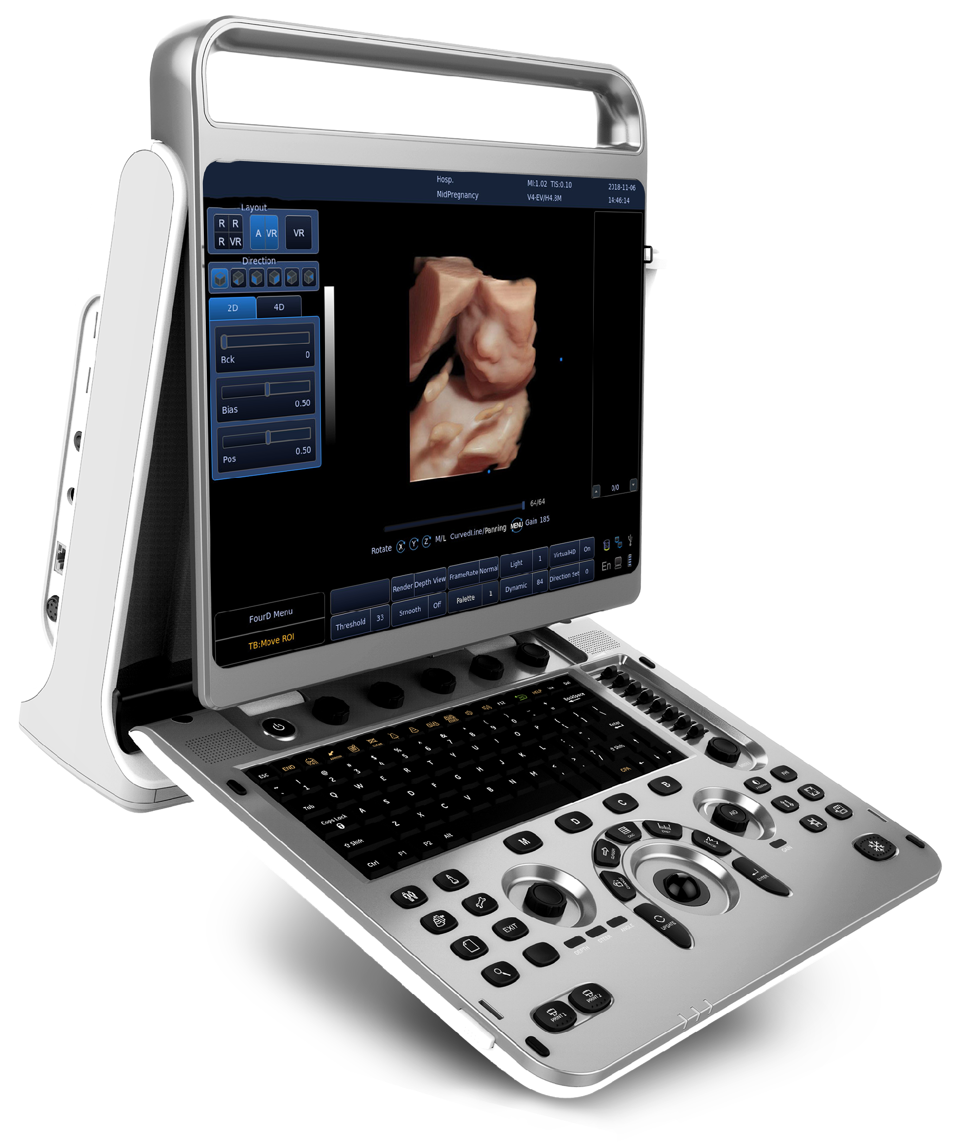
15 Inch Portable Color Doppler Ultrasound |
Application
· Abdominal
· Small organ
· Peripheral Vascular
· Transvaginal
· Transrectal
· Musculo-skeletal
· Pediatric
· Fetal
· OB
· GYN
· Urology
Feature
· FHI
-FHI is an innovative harmonic imaging technology that uses multiple transmission and receiving methods based on the patients' size and weight. This allows the EBit to maintain image resolution when imaging larger patients.
-Better than traditional THI and phased harmonic whichcompromise the penetration.
-FHI technology greatly improves diagnostic abilities and clinical confidence in larger, difficult-to-image patients.
· Q-image
-These innovative algorithms have strengthened the image enhancement results significantly.
-Advanced chipset is used to ensure fast frame rate.
· Q-beam
-Compared to the traditional dual-beam former on most ultrasound machines, the EBit 30 uses quad-beam technology for ultrasound signal receiving.
-Doubles the volume of signals received from traditional methods, increasing image resolution and generating more accurate images.
-Produces higher frame rates, ensuring better diagnostic confidence and efficiency, especially for moving organs.
· Virtual HD
-The latest innovation in real-time 4D with powerful imaging engine.
-Greatly strengthen the bond between mother and fetus.
-With moveable virtual light source.
· Elastography
-Elastography displays tissue stiffness in real time to provide doctors with additional diagnostic information when scanning organs like liver and breast.
Technical specification
1. Dimensions and Weight
Dimensions of main unit (approx.): 358mm(Length)×125mm(Width)×399mm(Height)
Net weight of main unit (approx.): 7.8kg (no probe included)
2. Electrical Power
Power supply voltage: Auto adaptable for AC100-240V
Power supply frequency: 50-60 Hz
Power consumption: 150 VA
3. User interface
Operation Panel
Control panel
Alphanumeric keyboard
8 STC Slides
Interactive backlit keys
High resolution color LCD
-Diagonal dimension: 15 inch
-Resolution: 1024X768
-Brightness adjustment
Integrated speaker
-Volume adjustable
4. Scanning Method
Electronic convex
Electronic linear
Electronic micro convex
5. Transducer Types
Convex transducer: C3-E
Linear transducer: L7-E, L12-E, L7W-E,
Transvaginal transducer: V6-E, V7-E
TransRectal transducer: L7R-E
Micro-convex transducer: MC3-E, MC6-E, MC5-E
6. Image Modes
B Mode
B/M Mode
M Mode
2B Mode
4B Mode
2D Steer (option)
CFM Mode
CPA Mode
DPD Mode
PW Mode
B/BC Mode (option)
Triplex Mode
Trapezoidal Mode
Super Needle (option)
Elastography Mode (option)
ECG (option)
7. Display Mode
Quad/dual display (Only for B)
Duplex mode: B+CFM, B+CPA, B+DPD, B/M
Triplex mode: B+CFM+PW, B+CPA+PW, B+DPD+PW,

Image Gallery:

Contact Information
E-mail:
Tel:
Address : Room 2512, Unit 1, Huaqiao Square, No.1, Daqiao North Road, Jiangbei New District, Nanjing, China
Veterinary Equipment
Medical Equipment

 There is currently no content to display


